This content is limited to our registered members
-
Access exclusive business services unlimited
-
Access valuable features : audio listening & tools sharing with your patients
-
Access more than 150 product sheets, dedicated to professionals
You already have an account ? login now
BIODERMA Congress Reports ESPD 2023
Reports written by Prof. Ivelina Yordanova (Dermatologist, Bulgaria) and Dr. Suzana Ožanić-Bulić (Dermatologist, Croatia)
Related topics
- Dryness / Atopy / Eczema
Today, 04 May 2023, we attended a remarkable Genodermatoses session on the programme of the 22nd ESPD Congress, held in sunny and welcoming Malaga in southern Spain, Andalusia region. In this session, Prof. John McGrath, Prof. Christina Haas and Dr. Angela Hernandez-Martin presented the latest developments in the genetic diagnosis, clinic and treatment of rare congenital skin diseases.
Speakers: John McGrath, Cristina Has and Angela Hernández-Martín
Report written by Prof. Ivelina Yordanova
New genodermatosis discoveries
John McGrath
Professor John McGrath, head of St John's Institute of Dermatology, King's College, London, professor of molecular dermatology and lead of the Genetic Skin Disease Group, presented the latest discoveries in the field of new genetic mutations and previously undescribed genodermatoses. He said that discovering the genetic basis of inherited skin diseases is fundamental to improving diagnostic accuracy and genetic counseling. Over the past decade the advent of next‐generation sequencing (NGS) technologies has accelerated diagnostic discovery and precision. The number of discoveries increased from approximately 40 disease‐associated genes prior to the identification of CYBB in 1986 to having over 1000 disease genes documented in the Online Mendelian Inheritance in Man (OMIM) database is currently available. One of the major promises of the genomic medicine era has been to give patients with inherited diseases accurate clinical diagnoses based on individual gene and mutation data. Implicit to this is the consideration of the relationship between individual genotypes and their associated clinical phenotypes. Of the 166 new disease–gene associations for inherited skin diseases discovered using NGS approaches between 2009 and 2019, there was an approximately even split between autosomal dominant and autosomal recessive conditions (51·2% vs. 47·0%, respectively), with the autosomal dominant conditions also divided approximately equally into dominant and de novo dominant inheritance. Other studies have similarly demonstrated the relative over‐representation of genes underlying autosomal recessive disorders reflecting the more straightforward filtering of homozygous or compound heterozygous variants compared with the identification of single and clearly pathogenic heterozygous variants for autosomal dominant diseases. From a clinical standpoint, the recognition of multiple molecular diagnoses can have important implications for genetic counseling, allowing more precise management and estimates of familial recurrence risk.
Genodermatoses: what can we treat?
Cristina Has
Prof. Cristina Has is a professor at the Department of Dermatology of the University of Freiburg in Germany, Head of the Molecular Dermatology Laboratory with a focus of interest on Epidermolysis bullosa and other genetic skin disorders. Her clinical activity includes general and pediatric dermatology, and genodermatoses. With her group, she has identified new genes and characterized large cohorts of patients with genodermatoses, established genotype-phenotype correlations and explored the underlying disease mechanisms. In the session today she presented what can we treat genodermatoses.
Due to the lack of effect and high toxicity of the treatment of some genodermatoses with classic drugs such as systemic retinoids, methotrexate and cyclosporine, we are turning our attention to new biological drugs, monoclonal antibodies blocking various molecules of the skin inflammation cascade. In addition to discovering genetic-clinical correlations, we are increasingly looking for pathogenetically-based therapeutic options targeting primary or reactive dysregulation. It turns out that different genetic defects lead to and trigger different tissue or system reactions related to skin inflammation and releasing of inflammatory cytokines - TNF alpha, IL-1, IL-17, IL-6, etc. The use of various biological molecules for treatment, significantly improves the quality of life of patients with genodermatoses and their families. As examples, she therefore indicated cases of a very good therapeutic effect achieved as a result of treatment of Epidermolysis bullosa dystrophica pruriginosa, which has a Th2 immune profile with biological drugs for the treatment of atopic dermatitis from the group of JAK kinase inhibitors - upadacitinib, baricitinib, tofacitinib and anti IL-4 - Dupilumab. Prof. Has presented a case of successful treatment of a patient with recessive dystrophic epidermolysis bullosa with Dupilumab in two different dosage regimens 200 mg/2 weeks and 300 mg/4 weeks. She also said that the drug losartan has an antifibrotic effect in cases of recessive dystrophic epidermolysis bullosa by reducing blistering, reducing esophageal strictures and restenosis, but not in all patients it has an effect. A change in the treatment model /paradigm/ of keratinization diseases, which have a complex immune profile related to an allergic response, similar to atopic dermatitis and psoriasis with predominance of IL-17 was shown. In fact, in the group of ichthyoses, the established primary genetic defects in the barrier structure of the stratum corneum lead to impairment of the protective function of the skin and an increase in its permeability to allergens, bacteria and viruses. This further increases skin inflammation. Due to this fact, a remarkable effect with reduction of erythema and pruritus was shown in the treatment of patients with Netherton's syndrome with anti-IL-17, anti-IL-12/23, anti-TNF alpha. Prof. Has presented cases of 6 children with Netherton's syndrome under 6 years of age successfully treated for erythema and pruritus with Dupilumab at a dose of 300 mg/4 weeks.
The result of systemic therapy with Gentamicin inducing protein expression in patients with specific stop codon mutations such as epidermolysis bullosa, Nagashima type palmoplantar keratosis, congenital hypotrichosis simplex of the scalp, was shown. The results of a Phase III clinical study approved by the EMA in the treatment of wounds in patients with junctional and dystrophic epidermolysis bullosa with Oleogel S10 - birch bark extract, were presented. Prof. Has also presented the current status of gene therapy in epidermolysis bullosa - ex vivo gene and stem cell therapy and in vivo gene therapy for dystrophic epidermolysis bullosa awaiting FDA approval.
Clinical practice and Genetics: a match made in heaven
Angela Hernández-Martín
Dr. Ángela Hernández-Martín, Senior Consultant in Department of Dermatology, Hospital del Niño Jesús, Madrid, Spain, secretary of the Spanish Group of Pediatric Dermatology, co-authored numerous chapter textbooks (including Bolognia ́s General Dermatology Textbook, Harper ́s Pediatric Dermatology Textbook, last editions) with main areas of interests keratinization disorders and neurocutaneous diseases, presented clinico-genetic correlations in ichthyoses and in particular several rare cases of ichthyosis with confetti.
Speakers: Maya El-Hachem and Iria Neri
Report written by Prof. Ivelina Yordanova
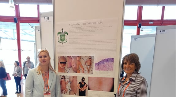
Neonatal skin signs suggesting genodermatoses
Maya El-Hachem
Today we attended a remarkable session on neonatal dermatology. The session was chaired by Dr. Maya El Hachem, a pediatric dermatologist at Bambino Gezu Children's Hospital in Rome.
She gave a very didactic lecture on "Neonatal skin signs suggesting genodermatoses". The group of keratinopathic ichthyoses was presented, which occurs with symptoms of congenital erythroderma and desquamation. Netherton's syndrome is characterized by congenital erythroderma, sparse hair, trichorexis invagina, atopic diathesis, and an elevated level of immunoglobulin E. It is associated with a complete absence of the LECTI protein, which is due to a mutation in the SPINK5 gene. Desquamation and hyperkeratosis in the neonatal period are signs of ichthyoses, representing a group of genodermatoses characterized by congenital damage to keratinization. An example of the most severe ichthyosis is Harlequin-ichthyosis. Restrictive dermopathy is a rare genodermatosis due to ZMPSTE 24, more rarely to LMNA mutation. This disease is characterized by an extremely poor prognosis, resulting in early neonatal death. Clinical signs are fetal transfer, intrauterine fetal retardation, thin rigid skin with lacerations in the folds, facial dysmorphism, and joint ankyloses. Incontinentia pigmenti is a genetic disease associated with a mutation of NEMO gene located on the X-chromosome.
Mosaicism due to lionization is common here. It affects only the female sex, it is fatal for the male and characterized by diffusely located vesicles along Blashko's lines, which subsequently turn into hyperkeratotic lesions, healing with postlesional linear hypopigmentation. It is associated with extracutaneous signs. Epidermolysis bullosa congenita is a clinically and genetically heterogeneous group of genodermatoses characterized by skin and mucosal blistering after minimal trauma. They are inherited in an autosomal dominant or recessive manner. There are 4 main variants - simplex, junctional, dystrophic and Kindler syndrome. All EB subtypes are caused by mutations in 16 genes encoding total of 13 proteins of the dermo-epidermal membrane zone structure. Skin atrophy and poikiloderma are typical of Kindler syndrome. Tuberous sclerosis is an autosomal-dominant disease characterized by multiple dysplastic organ lesions and neuropsychiatric symptoms. Mutations are present in 70% of patients and 2/3 have occurred daily. Congenital giant melanocytic nevi are associated with genetic mutations in the NRAS gene, which disrupt the normal differentiation and proliferation of melanoblasts. Patients with this diagnosis should be followed up by MRI for neuromelanosis. Clinical and dermatoscopic follow-up should be performed. In conclusion, neonatal skin manifestations can predict a wide and heterogeneous group of genodermatoses. Diagnostic tests, including molecular genetic testing, should be performed as early as possible. Multidisciplinary management is mandatory to ensure adequate care. Genetic counseling should be provided for the parents and psychological support should be provided.
Erythroderma in the newborn
Iria Neri
The second presentation in this short session was on neonatal erythroderma and was presented by Dr. Iria Neri. Erythroderma is a persistent generalized skin erythema affecting at least 90% of the skin surface. Neonatal erythroderma can appear already during birth or in the first 4 weeks after it. Depending on the degree of epidermal disturbance, erythroderma can cause serious complications such as electrolyte imbalance, hypoalbuminemia, dehydration, temperature instability, infections that could even lead to sepsis. In the neonatal period, erythroderma can be a manifestation of a number of congenital conditions. Dr. Neri presented the 6-step approach to the diagnosis of the relevant genodermatosis, published in 2022 in JEADV. Neonatal erythroderma is also rare - only 74 cases were identified in a 30-year period. Diagnosis is often a challenge for clinicians and is usually delayed due to the nonspecific nature of clinical signs. Clinical clues and diagnostic tests can be of great importance in making the final diagnosis. Three are the most common diseases occurring with neonatal erythroderma, accounting for 64% of cases. These are the ichthyoses, Netherton syndrome and Omenn syndrome. Omenn syndrome is characterized by the following clinical signs: pachydermal thickening of the skin, alopecia. Parents are usually consanguineous. Extracutaneous signs include severe growth failure, lymphadenopathy, diarrhea, infections, hepatosplenomegaly. The term Collodion baby describes a number of syndromes, in 75% of cases it is represented by autosomal recessive congenital ichthyoses, in 10% - self-healing Collodion baby and in 15% - other keratinizing diseases. In conclusion, neonatal erythroderma is rare, in the neonatal period it may be the first manifestation of many diseases whose typical clinical phenotype may appear later.
Speaker: Lisa Weibel
Report written by Prof. Ivelina Yordanova
The last day of the Congress of the European Association of Pediatric Dermatology 06/05/2023 in Malaga was full of new information about the latest clinical studies in the field of pediatric dermatology. We are now in the golden years of pediatric dermatology for the treatment of a number of childhood widespread and rarer childhood diseases. The reason is that we live in the era of biologicals, which have rapidity of action and high efficiency. Targeted therapies have almost minimal risk of organ damage. The opportunities for gene therapies in genodermatoses are increasing. We are still working with conventional systemic treatment. The choice of therapy requires an in-depth knowledge of the benefits and risks of each drug in patients, each of whom is an individual case.
An exceptional lecture was given by Dr. Lisa Weibel, head of the Department of Pediatric Dermatology at the University Children's Hospital Zurich, on the topic "Skin manifestations of systemic diseases". Dr. Weibel has two medical specialty - in pediatrics and dermatology, making her an exceptional specialist in the field of pediatric dermatology.
The skin is not an isolated organ, it participates in all processes that occur in the human body. When examining a child, we must look closely at the skin and skin appendages, fingernails and toenails, hair, where we may find some signs of internal, sometimes life-threatening diseases. She provided an in-depth review of skin manifestations in genetic disorders, conditions associated with developmental abnormalities, infectious diseases, immunologic inflammatory diseases, and childhood malignancies. Nail patella syndrome is an autosomal-dominant inherited genetic disorder characterized by progressive nail dystrophy from infancy, starting initially in the fingers and subsequently affecting the toes, presenting with severe dystrophy , hypoplasia and longitudinal striae or splitting of the nail plates. Nail dystrophy is the first sign that may lead to suspicion of nail patella syndrome, characterized by dysplastic or missing patella, glaucoma and impaired renal function in 30-50% and terminal renal failure in 15% of cases. Erythropoietic protoporphyria, inherited in an autosomal recessive manner, results in the absorption of visible light by protoporphyrin, leading to capillary damage. Indications for this disease can be skin manifestations in the child's preschool age, expressed in painful swellings and erythema of the hands, ears, nose, covered with crusts after sunny days. Mild liver damage is usually found in the disease, in 5% - liver failure. Degos disease or malignant atrophic papulosis is an extremely rare disorder in which small and medium sized arteries become blocked (occlusive arteriopathy), restricting the flow of blood to affected areas. Degos disease usually causes characteristic skin lesions that may last for a period of time ranging from weeks to years - "porcelain" shining atrophic papules on the eyelids, enlarged nail folds capillaries. Children diagnosed with this disease may develop complications due to impairment of internal organs. Similar papules can also form in patients with Dermatomyositis, but a different histopathological picture there is established. Acute leukemia is not a rare disease in children and is always characterized by high fever and erythematous macules on the body. The diagnosis is confirmed immunohistochemically, by taking a skin biopsy and must be made in time in order to start a specific treatment which is a bone marrow transplant. With long-term use of Levamisole for the treatment of nephrotic syndrome in childhood, in some cases, circulating autoantibodies (antinuclear, antiphospholipid and anticytoplasmic) are formed, which lead to necrotizing vasculitis and vasculopathy, manifested clinically as purpura of the ears, nose and auricles. These skin changes always resolve spontaneously when treatment with Levamisole is discontinued. The appearance of ecthyma-like necrotic changes on the skin of the torso immediately after birth is a sign of congenital Langerhans histiocytosis. In the presence of epidermolysis of the skin at birth, as well as the appearance of bullae and desquamation of the palms and soles, in addition to congenital epidermolysis bullosa, epidermolytic ichthyosis and SSSS syndrome, we must also think about congenital syphilis and perform the relevant serological tests. In congenital syphilis, mucosal involvement is observed in up to 60% of cases. The presence of cutaneous granulomas from infancy is associated with primary immunodeficiency diseases. Oculocutaneous albinism, bleeding diathesis, Crohn's-like enteropathy and pulmonary fibrosis are associated with Hermansky-Pudlak syndrome, which is a very rare autosomal recessive genodermatosis. A skin manifestation of Hodgkin's Lymphoma, which represents 7% of childhood neoplasias affecting the age of 14-19 years, is persisting pruritus (eczema) in 20-50% of cases. When eczema started at this age, it is appropriate to refer patients for MRI and CT of the lung, where enlarged mediastinal lymph nodes can be detected. Evaluation of multiple atypical cafe-au-lait-spots. When they are typical and combined with axillary freckling, it is Neurofibromatosis 1. When they are atypically located, they are associated with increased tumor formation in early childhood. When the spots are hypopigmented, signs of Fanconi anemia type D1 should be looked for, because these patients carry a genetically high risk for the development of solid tumors - medulloblastoma, Wilms' tumor, neuroblastoma, acute leukemia, before the age of 5 years. Strict tumor screening is required in these patients. The management of small patients with skin manifestations of life-threatening diseases requires a multidisciplinary approach and collaboration between dermatologists, rheumatologists, pediatricians, neurologists, oncologists, immunologists and endocrinologists.
Dear colleagues,
It was a pleasure and honour to participate at 22nd ESPD meeting in Malaga. Apart from excellent lectures, meeting colleagues from different countries was a fantastic opportunity to exchange ideas and share knowledge and experience from paediatric dermatology. Please find below report from lectures that were of special interest to my daily practice.
Speaker: Kinsler Veronica MD, Professor of Paediatric Dermatology and Dermatogenetics
Report written by Dr. Suzana Ožanić-Bulić
1. What drives the melanoma risk in children with CMN?
- CMN are most commonly caused by mosaic mutations affecting NRAS or BRAF
- Primary melanoma in children with multiple CMN arises most commonly in the central nervous system
- Melanoma risk is therefore unlikely to be strongly related to UVR (ultraviolet radiation) and more related to the genetics
- For this reason, only standard good sun protection measures are advocated
2. What is the Vitamin D status in children with CMN and can we recommend what to do?
- 40% of CMN patients in the UK cohort had Vitamin D insufficiency or deficiency
- 27% of levels were insufficient (30-49 nmol/L), 13% deficient (<30 nmol/L) by age-matched UK standards
- Total vitamin D levels were not statistically significantly influenced by age, sex, CMN projected adult size, or season in which the sample was taken (regression analysis)
3. How does this compare to the general UK population?
- Figures are comparable to those in general European populations
- Current UK National Institute of Clinical Excellence (NICE) guidelines are
- Deficiency: high-dose Vitamin D followed by daily maintenance
- All individuals should consider taking 10 micrograms of Vitamin D daily all year
- At-risk children should take 10 micrograms Vitamin D daily all year
4. Conclusions for the UK
- Sun protection for children with CMN should be (excellent and) the same as for all children
- Children with CMN should have 10 mcg of Vitamin D orally daily, at least September-April each year
- Routine serum measurement of Vitamin D is not required
Speaker: Ott Hagen, Professor of Paediatric Dermatology and Allergology
Report written by Dr. Suzana Ožanić-Bulić
1. Classification of AIBD (Autoimmune Bullous Diseases)
- Subepidermal
- Linear IgA bullous dermatosis
- Bullous pemphigoid
- Epidermolysis bullosa acquisita
- Dermatitis herpetiformis
- Intraepidermal
- Pemphigus vulgaris
- Pemphigus foliaceus
- Neonatal pemphigus
- Paraneoplastic pemphigus
- Endemic pemphigus
2. AIBD - differential diagnosis includes various infectious diseases, hypersensitivity reactions, hereditary diseases and trauma
- Bacterial and viral infections
- Bullous impetigo
- Staphylococcal scalded skin syndrome (SSSS)
- Herpes simplex infection (HSV)
- Enterovirus
- Hypersensitivity reactions
- Stevens Johnson Syndrome (SJS)/Toxic Epidermal Necrolysis (TEN)
- Mycoplasma pneumoniae induced rash and mucositis (MIRM)
- Bullous EEM (Erythema exudativum multiforme)
- Hereditary diseases
- EB (Epidermolysis bullosa) hereditaria
- Epidermolytic ichthyosis
- Peeling skin syndrome
- Trauma
- Dermatitis artefacta
- Thermal injury
- Insect sting reaction
- Others
- Bullous mastocytosis
- Bullous scabies
- Phototoxic reactions
3. AIBD - diagnostic Work-up. No effective and safe treatment without histology, DIF, ELISA…)
- Clinical features (AIBD is not a clinical diagnosis!)
- Histology
- DIF (Direct immunofluorescence)
- IIF (Indirect immunofluorescence)
- Autoantigens (DSG1, DSG3, BP 180, BP 230, LAD-1, Collagen VII…)
4. AIBD – think of multisystem disorders
- Impaired homeostasis – dehydration, electrolyte imbalance, hypothermia
- Severe skin symptoms – debilitating pruritus, pain, erosions, ulcerations, blistering, exfoliation
- Increased risk of infections - bacterial skin infections, viral skin infections, systemic infections, septicaemia
- Psychosocial stress – stigmatization, school absence, anxiety (patient, parents, siblings), therapy costs
- Extracutaneous complications – oesophageal strictures, drug side effects, IgA nephropathy
- Failure to thrive – oral blistering/ulcerations, feeding difficulties, higher energy reauirements
5. Multiprofessional care most important in effective treatment outcomes
- Wound care team
- Pain management
- Psychosocial support
- Ophthalmology
- Otolaryngology
- Gastroenterology
- Paediatrics
- Dermatology/Paediatric dermatology
6. Children with AIBD should be vaccinated
2022 American College of Rheumatology Guideline for Vaccinations in Patients with rheumatic and musculoskeletal diseases
- Pneumococcal vaccine for all individuals under immunosuppression
- Continuing immunosuppressive medication around time of non-live attenuated vaccinations
- Hold immunosuppressants before live-attenuated vaccination (shown in table with timeline for each immunosuppressant with specific periods before starting the treatment and time required after treatment was stopped)
7. Therapeutic desert – no blinded randomized controlled trials
- Therapeutic armamentarium
- Topical and systemic glucocorticosteroids
- Dapsone
- Doxycycline
- Methotrexate
- Mycophenolate
- Azathioprine
- IVIG
- Rituximab
8. Subepidermal AIBD topical treatment
- Topical anti-inflammatory treatment – methylprednisolone 0.1%, mometasone 0.1%, clobetasol 0.05%
- Tapering schedule – month 1: daily treatment, month 2: every 2nd day, month 3: 2 times per week, month 4: once per week
9. Subepidermal AIBD systemic treatment
- Systemic corticosteroids
- Start: prednisone 0.5 mg/kg/d
- If no control of disease activity (CDA) on day 15: 0.75 mg/kg
- CDA: slow tapering (e.g. 25% dose reduction weekly/biweekly)
- Goal: minimal therapy (0.1 mg/kg/d)
- Dapsone
- Start with 0.5 mg/kg/d
- Increase to 1(-2) mg/kg/d
- Lab: methaemoglobin, CBC/DIFF
- Intravenous immunoglobulins
- 2 g/kg every 4 weeks
- Mycophenolate mofetil
- 300 mg/m2 twice daily
- Emerging therapies: Biologics
10. Emerging therapies in BP (Bullous pemphigoid) – case reports
- Two cases of BP effectively treated with oral tofacitinib
- Bullous pemphigoid treated with Janus kinase inhibitor upadacitinib
11. Pemphigus vulgaris treatment
- 1st line treatment
- Rituximab two infusions of 1g two weeks apart + systemic corticosteroids (prednisone 1mg/kg/day) with a progressive tapering in order to stop corticosteroids after 6 months or
- Systemic corticosteroids oral prednisone 1 to 1.5 mg/kg/day alone or associated with an immunosuppressive drug (azathioprine 1 do 2.5 mg/kg/day or mycophenolate mofetil 2g/day or mycophenolate sodium 1440 mg/day)
Speaker: El Hachem May MD, Head Paediatric Dermatology Unit
Report written by Dr. Suzana Ožanić-Bulić
1. Genodermatoses: neonatal skin signs
- Erythroderma
- Scaling/hyperkeratosis
- Vesicles/blisters
- Plaques/nodules
- Dyschromic lesions
2. Erythroderma – erythematous cutaneous eruption characterized by
- Skin involvement over 90%
- Severity
- Chronic evolution
- Frequent associated scaling
- May suggest rare and common diseased - Ichtyosis, immunodeficiency, metabolic disorders, psoriasis, atopic dermatitis, seborrheic dermatitis
3. Keratinopathic ichthyoses
- Erythroderma with desquamation
- Superficial blistering
- Comprise: epidermolytic ichthyosis, ihthyosis with confetti, superficial epidermolytic ichthyosis, and epidermolytic naevus
- Due to mutation in keratin genes
4. Netherton syndrome
- Erythroderma
- Hypernatremia
- Sparse hair
- Trichorrhexis invaginate (not present early in life)
- AD and hyper IgE
- Diagnosis: complete lack of LEKTI, mutation in SPINK5
5. Scaling/hyperkeratosis
Ichtyoses – heterogenous group of genodermatoses characterized by hereditarian disorder of keratinization defect of maturation of the basal layer cells and scaling
6. Restrictive dermopathy
- Rare genodermatosis, caused by ZMPSTE24, or less frequently, LMNA mutations
- Characterized by extremely poor prognosis resulting in stillbirth or early neonatal death
- Clinical features comprise prematurity, intrauterine growth retardation, thin rigid skin with lacerations at flexural regions, facial dysmorphism and joint ankyloses
7. Incontinentia pigmenti
- Genetic disorder
- X-linked (Xq28) – NEMO gene
- Mosaicism due to lyonization (random inactivation of one chromosome X during embryology)
- It affects female (usually lethal in male)
- Characterized at birth by diffuse vesicles following Blaschko lines
- Association with extracutaneous manifestations
8. Epidermolysis bullosa
- The most recent classification distinguishes 4 major types following the cleavage level
- EB Simplex, within basal keratinocytes
- Junctional EB, within the lamina lucida
- Dystrophic EB, below the lamina densa
- Kindler EB, mixed levels
- EB causative genes and encoded proteins – EB subtypes are caused by mutations in 16 genes encoding 13 proteins
9. Aplasia cutis in EB
- Congenital skin defects usually affecting the lower limbs
- Described in all EB types, more frequently in EBS intermediate with cardiomyopathy (KLHL24) and dystrophic EB, in particular dominant intermediate and self-improving subtypes (COL7A1)
10. Skin atrophy in EB
- Skin atrophy and poikiloderma are typical of Kindler EB (FERMT1)
- Skin atrophy can manifest already in infancy
11. Tuberous sclerosis
- Tuberous sclerosis complex (TSC) is autosomal dominant disorder characterized by multiple dysplastic organ lesions and neuropsychiatric symptoms, caused by loss of function mutations in either TSC1 or TSC2 gene
- Mutations are present in 70% of patients, and 2/3 are de novo mutations
12. Dyschormic lesions: pigmented and vascular
Giant congenital melanocytic naevi (GCMN)
- Rare
- Usually associated with genetic mutations in NRAS (<KRAS) that interfere with the normal proliferation, differentiation and migration of melanoblasts
- Diagnosis – brain and spine MRI to investigate neuromelanosis
- Clinical and dermoscopic follow-up
- Nodules within GCMN may be – proliferative and benign or melanoma
13. Conclusion
- Neonatal skin manifestrations may be suggestive of a wide and heterogenous group of dermatoses
- Diagnostic workup, including molecular genetic testing, should be started as soon as possible
- Multidisciplinary management is mandatory to guarantee an appropriate care
- Genetic counselling should be offered to the parents
- Psychological support is necessary
Speaker: Baselga Eulalia MD, Professor of dermatology
Report written by Dr. Suzana Ožanić-Bulić
1. Vascular stains in the newborn: pattern recognition
- Is it vascular?
- Is it vascular malformation or a tumour?
- Do we need workup?
2. Is it vascular?
- Colour
- It blanches
- Shape and localization
- No textural skin changes
3. Is it vascular malformation or tumour?
- Precursor of haemangioma (white peripheral halo) – diagnostics for PHACE
- Capillary malformation (port wine stain) – diagnostics for SWS
- Stain of an AVM (arterio-venous malformation) or a RASA1 stain – Arteriography
4. PHACEs (OMIM 606519) – 30-46% segmental IH face and scalp
- Posterior fossa malformation
- Large segmental Facial Haemangioma
- Arterial anomalies
- Cardiac and Coarctation
- Eye anomalies
- Sternal raphe
- Work-up: MRI with contrast and Angio MR head and neck, Echocardiogram, Ophthalmologist referral, Hearing test
5. Propranolol in PHACE
- Cardiac contraindication… only if bradycardia
- Echocardiogram including aorta, start propranolol at lower dose (0.5 mg/kg) in three divided doses
- Theoretical risk of hypoperfusion
- Stroke PHACE and propranolol: no evidence
6. Sturge-Weber Syndrome (10-35% PWS forehead area)
- Leptomeningeal angiomatosis (LMA)
- Early onset seizures
- Stroke-like episodes
- Neurodevelopmental delay
- Glaucoma
- Risk of SWS in frontal PWS – all MRI before 1 year, 16% had SWS
7. Patients at risk SWS
- Ophthalmology referral
- MR with gadolinium “gold standard”
- ECG in specialized centre
- Workup necessary – parents want to know, parents can be trained to recognize often subtle first seizures, early treatment can prevent generalization or status epilepticus and neurodevelopmental outcome, preventive treatment in extensive cases
8. Early MRI: At what age?
- LMA visualisation needs gadolinium
- False negative results before 2-3 months
- Need for anaesthesia (“Feed and wrap” technique)
- Gadolinium toxicity (unheard in infants and with newer macrocyclic compounds)
9. is it vascular?
- Stain of an AVM
- Warm to palpation
- Rapid capillary refill
- White peripheral halo
- Colour heterogeneity
10. “Dominant” stain of CM-AVM: Do we need workup?
- Autosomal dominant, hight penetrance
- Germline loss of function in RASA-1 (50%), EPHB4 (25%), mosaic RASA-1
- 1/3 AVM: 10% intracranial AVM – haemorrhage, high output heart failure
11. Vascular stain on lower lip
- Pattern recognition – shape is characteristic stain of CLAPO
- PIK3CA mutation
- +/- lymphatic malformation
- +/- overgrowth
12. Take home messages
- PAST – pattern recognition, descriptive names
- NOW – molecular characterization, genotype-phenotype correlation, rapamycin for everything, few targeted therapies
- FUTURE – biomarkers, risk stratification of patients, evidence-based recommendations
Speaker: Mulleger Robert MD, Prof dermatologist
Report written by Dr. Suzana Ožanić-Bulić
1. Lyme Borreliosis (LM)
- Multisystemic infectious disease
- Borrelia burgdorferi (Bb) sensu lato
- Transmission by hard ticks (family: Ixodidae)
- Most frequent tick-transmitted disease in northern hemisphere
- Incidence peaks: 5-15 ys and >50 ys
- Same spectrum in children and adults
- Differences in prevalence and clinical characteristics
2. Stages of LB and clinical manifestations
- Early localized
- Erythema migrans
- Borrelial lymphocytoma
- Disseminated EM
- Early disseminated
- Disseminated EM
- Aseptic meningitis
- Mono/polyradikuloneuritis
- Cranial nerve palsies
- Encephalitis, myelitis
- Arthritis
- Myositis
- AV-block I-III, myopericarditis, endocarditis, pericarditis, tachycardia
- Conjunctivitis, iritis, (chorio)retinitis, uveitis, keratitis, optic nerve neuritis
- Late
- Acrodermatitis chronica atrophicans (ACA)
- Peripheral (poly-) neuropathy
- Chronic encephalomyelitis
- Cerebral vasculitis
- Chronic arthritis
- Dilated cardiomyopathy
- Keratitis
3. Erythema migrans
- Hallmark of early LB
- Single or multiple, expanding, round-to-oal, bluish-red, sharply demarcated erythema(s) of at least 5 cm
- Either ring-like with central clearing or homogenous
- 45% of all LB manifestations in children (>70% in adults)
4. Borrelial lymphocytoma
- Early localized, subacute skin manifestation of LB
- One of the most common B-cell pseudolymphomas
- Almost exclusively in Europe
- 50% of cases in children
- Non-tender, soft, bluish-red nodule or plaque of 1-5 cm
- Site in children: ear (88%), breast (7%), scrotum (7%)
5. Acrodermatitis chronica atrophicans
- Cutaneous manifestation of late stage LB
- Extensor surfaces of (distal) extremities
- Inflammatory stage evolves into atrophic stage over months
- Rarest form of LB, especially in children
6. Therapy of LB in childhood – antibiotics
(oral therapy to be preferred, except for severe neurologic or cardiac manifestations)
- Amoxicillin p.o., 50 mg/kg (max 1.5 g) in 3 divided doses
- Cefuroxime axetil p.o., 30 mg/kg (max 1g) in 2 divided doses
- Azithromycin p.o., 10 mg/kg (max 500 mg) once daily (5-10 days)
- Phenoxymethyl penicillin p.o., 200.000 – 400.000 U/kg (max 18-24 Mio. U) every 4 hrs
- Doxycycline p.o., 4 mg/kg (max 200 mg) in 2 divided doses – contraindicated in children <8 ys
- Low risk of permanent tooth staining or enamel hypoplasia (scarce data)
- Ceftriaxone i.v., 50-75 mg/kg (max 2g) in one dose
- Duration of therapy
- Early localized disease 10-14 days
- Early disseminated disease 14-21 days
- Late 14-28 days
7. Treatment outcomes
- Excellent outcome after antibiotics
- Prompt clearance of skin lesions
- EM + MEM: med. <1 wk- 2 ws (max 3 months)
- BL: med 2 ws – 2 months (max 10 months)
- Extracutaneous symptoms disappear within a few days
- Persistence of headache a/o arthralgias for max 3 mo
- No late sequelae
- Jarisch-Herxheimer reaction rare
Speaker: Horev Amir MD
Report written by Dr. Suzana Ožanić-Bulić
Cutaneous Leishmaniasis
- No perfect treatment in children – painful, irritable, not approved for young children, drug toxicity, not suitable for all Leishmania species, scarce data regarding clinical efficacy, hight economic cost
- Topical
- Thermotherapy
- Cryotherapy
- Paromomycin
- Pentostam injection
- CO2 laser
- Liposomal amphotericin B
- Photodynamic patch
- Nitric oxide
- Oral
- Rifampin
- Fluconazole
- Itraconazole
- Ketoconazole
- Azithromycin
- Miltefosine
- Allopurinol
- Parenteral
- Pentostam
Speaker: Theiler Martin MD, Consultant for Paediatric dermatology
Report written by Dr. Suzana Ožanić-Bulić
1. Unusual exanthems – case presentations
- Infant with fever and rash
- JXGs followed by an unusual exanthem
- DRESS or no DRESS
2. Case presentation – infant with fever and rash
- 9 mo infant with fever and maculopapular exanthem for 3 days, conjunctival injection, erythematous and scabby lips, cervical lymphadenopathy, CRP 85 mg/L and microcytic anaemia
- Differential diagnosis – Kawasaki disease, COVID -19 (MIS-C, PIMS), Toxic shock syndromes, Measle or other viral exanthems, Drug reaction (DRESS)…
- Diagnosis Febrile ulceronecrotic Mucha-Habermann disease (FUMHD)
- Acute and severe variant of pityriasis lichenoides with systemic involvement
- High fever, gastrointestinal symptoms, CNS manifestations, pulmonary involvement, cardiomyopathy, pancytopenia, DIC, sepsis
- Infectious trigger in a proportion of cases
- Males predominantly affected
- Mortality 2% in children (20% in adults)
- Treatment – systemic steroids and methotrexate
3. Case presentation – JXG followed by an unusual exanthem
- 7 mo girl with multiple juvenile xanthogranulomas (JXG)
- Normal ophthalmology exam, normal abdominal ultrasound, normal differential full blood count, no evidence of neurofibromatosis
- New rash developed with differential diagnosis – Sweet syndrome, insect bite reaction, lymphomatoid papulosis
- Diagnosis skin rash – Sweet syndrome
- Postinlammatory elastolysis /cutis laxa – up to 30% of paediatric Sweet syndrome, associated with cardiovascular complications (aortic aneurysm, stenosis of coronary vessels…)
6. Case presentation – DRESS to amoxicillin or DRESS-like viral exanthem (+ amoxicillin)
- Lymphadenopathy, no eosinophilia, atypical lymphocytes (low number), transaminases 2 times ULN
- Early-onset antibiotic-induced DRESS – time of onset 7-19 days, eosinophilia +++, other organ involvement ++, significant improvement 3-5 weeks with corticosteroids, no evidence of viral infection
- DRESS-like rashes during viral infections and antibiotic intake – time of onset 6-7 days, no eosinophilia, no other organ involvement, significant improvement 2-5 days with/without corticosteroids, positive evidence for viral infection
7. Take home messages
- FUMHD may have Kawasaki-like clinical presentation and mucosal involvement
- Methotrexate is effective treatment for FUMHD
- Sweet sy is associated with malignancy in 25% of children
- 30% of children with Sweet sy develop elastolysis that causes cardiovascular complications
- Viral exanthems treated with betalactams may have a DRESS-like clinical presentation
- The lack of eosinophilia and the faster resolution differentiate them from true early-onset DRESS
Speaker: Wollenberg Andreas, Professor of Dermatology and Allergy
Report written by Dr. Suzana Ožanić-Bulić
1. A treatment for each phenotype
- Which phenotypes of AD do exsist?
- Which aspects should influence our treatment decision?
- Which problems should we solve?
- The framework of AD guidelines
- Topical treatment of AD
- The indication for systemic treatment
- Current options for systemic treatment
- Choice of treatment regimen
- Outlook and practical issues
2. Phenotypes of AD
Intertriginous AD, palmoplantar hyperkeratotic AD, heavily colonized AD, nummular AD, hyperxerotic AD, eczema herpeticum prone AD, flexural AD, impetiginized AD, palmoplantar dyshidrosiform AD, UV-light triggered AD, head-and-neck AD, exudative AD, AD in ichthyosis, Atopic eyelid dermatitis
3. Patient characteristics and problem to solve are different for each patient
- Acute flare of disease with need for acute intervention
- Long term chronic disease with a need for immune modulation
- Severe atopic distortion with a need to prevent flares
4. Treatment of atopic dermatitis – therapeutic options in 2022
- Step care plan for children and adolescents with AD
- Baseline therapy with emollients, avoiding of allergens, educational programmes
- Mild AD – baseline measures + TCS acute or TCI reactive + wet wraps acute
- Moderate AD baseline measures + TCS and TCI proactive, NB-UVB and medium dose UVA1, psychosomatic counselling
- Severe AD – baseline measures + systemic treatment: Cyclosporin A – in license for >=16 ys, dupilumab in license for >=6 ys, Upadacitinib in license for >=12 ys, Azathioprine and Methotrexate not licensed in children with AD
5. Epidermal barrier improvement
- Reduction of enzymatic SC degradation – adjustment of skin pH including Filaggrin adjustment, supplementation of Protease inhibitors
- Hydration of SC – glycerol and urea, stimulation of Filaggrin production / Filaggrin supplementation
- Supplementation of lipid lamellae constituents – ceramide containing products
- Physical measurements – short time rinsing, showering and bathing with syndets, soak and seal technique, no extensive rubbing
6. Wet wrap therapy with emollients and TCS
- Steroid sparing treatment of choice for very dry, moderately inflamed skin
- Acute intervention treatment for severe AD if used with diluted TCS
7. Emollients plus
- Emollients – topical treatment with vehicle-type substances lacking active ingredients
- “Emollients plus” – several non-medicated products for topical treatment of AD contained putative active ingredients (e.g. flavonoids, saponins and riboflavins…), but are neither fulfilling the definition of nor needing a licence as a topical drug
- Emollients containing potentially allergenic plant proteins from peanut, oat or wheat should be avoided in children before the age of 2, whereas protein-free plant extracts are apparently safe to us
8. Proactive therapy of AD (Tac and TCS)
- Defined as long-term, low-dose intermittent application of anti-inflammatory therapy to the previously affected skin together with an ongoing emollient treatment of unaffected skin
9. Indication for systemic treatment - candidates for systemic treatment
- Patients who have high composite score such as SCORAD >50 (Scale definition)
- Patients who are clinically not responding to an appropriately conducted topical therapy (Functional definition)
- Patients who are unable to participate in normal daily life activities while following an adequate treatment regimen (Social definition)
- A signs-only score, such as EASI, is not adequate tool to discriminate between providing and declining systemic therapy to an individual patient
10. TH2 blockers – Dupi, Tralo, Lebri
- Dupilumab – mAb against the alpha chain of the IL-4 and IL-13 receptor (blocking both signals)
- Tralokinumab – monoclonal antibody against cytokine IL-13
- Lebrikizumab – monoclonal antibody against cytokine IL-13
11. Tyrosin kinases in Atopic dermatitis
- Baricitinib, Upadacitinib, Abrocitinib
- Very common and common adverse reactions in AD across JAK inhibitors
- Each JAK inhibitor has its own safety profile
12. Drug-drug interactions profiles differ between JAK inhibitors
- Relevance for CYP450 drug interactions – yes for abrocitinib and upadacitinib, no for baricitinib
13. EMA: JAKi Article-20 referral
- JAK inhibitors should be used only if no suitable treatment alternatives are available in patients: aged 65 years or above, at increased risk of MACE, those who smoke or have done so for a long time in the past and those at increased risk of cancer
- JAK inhibitors should be used with caution in patients with risk factors for blood clots in the lungs and veins (venous thromboembolism, VTE) other than those listed above
- Doses should be reduced in some patient groups who may be at risk of VTE, cancer or major CV problems
- No changes were recommended to the currently approved indications for all JAK inhibitors
14. What will be the future?
- We should define better which clinical problem should be addressed by a systemic drug
- All licensed substance classes have key advantages and disadvantages
Speaker: Graaf Marlies MD, PhD, Paediatric dermatologist
Report written by Dr. Suzana Ožanić-Bulić
1. Need for treatment
- Impaired skin barrier
- Immune dysfunction
- Impact on quality of life (QoL) of patient and parents
- Increased risk for developing atopic comorbidities
2. Dual-allergen exposure hypothesis
- Cutaneous exposure – skin, skin-draining lymph nodes, Th2 memory, allergy
- Oral exposure – GI tract, mesenteric lymph nodes, Th1 memory, Treg memory, tolerance
- Prevention studies on early food introduction to prevent FA (LEAP, EAT, PETIT)
- Early introduction – worldwide recommendation: introducing peanut <12 months of age without previous testing appears to be safe and effective
3. Systemic treatment options for infants (<2 years)
- Conventional immunosuppressants: cyclosporin A, methotrexate, azathioprine, mycophenolate mofetil – off-label, unfavourable safety profile, lack of studies, less experience in infants
- New immunomodulating therapies: dupilumab, tralokinumab, upadacitinib
4. Dupilumab for AD in children >6 months-6 years
- Safety – conjunctivitis (5%), transient increase in mean eosinophil count, less skin infections
5. Challenges in treating infants with severe AD
- Practical challenges – indication for starting and stopping, subcutaneous injections
- Knowledge gaps – longterm effects, impact on comorbidities, effectiveness of vaccination, impact on globet cells
6. Take to work messages
- Treat atopic dermatitis
- It is not yet clear whether and how AD can be prevented
- Well-controlled AD may play a roll in the prevention of food allergies
- Important to properly treat AD and new systemic options will help in severe cases
- Atopic comorbidities
- Young infants with AD are particularly at risk of developing food allergies
- Early introduction of food allergens decreases the risk of developing food allergy
- Timing is essential (“the earlier the better”)
Speaker: Silverman Robert MD, Dermatologist
Report written by Dr. Suzana Ožanić-Bulić
1. Nail disease in paediatric dermatology practice
- 2% of office visits to paediatric dermatology clinic
- Chief complaints:
- Loss of nail plate
- Growths
- Fungus – blisters, pain
- Discoloration
2. Nail changes
- Endonyx
- Isolated 4th toenail onychomycosis
- Elkonyxis
- Parakeratosis pustulosa
- Epidermolysis bullosa
3. Childhood tinea unguium
- Prevalence 0.2-2.6% in developed countries
- Increased incidence – Down syndrome, HIV, children from households with moccasin type T. rubrum
- Clinical variants similar to adults
4. Endonyx
- Absent inflammation, onycholysis or subungual hyperkeratosis
- Undisturbed nail plate architecture, normal thickness
- Eventually affects the entire thickness of the nail plate – milky white colour, usually fingernails
- Trichophyton soudonense, violaceum, tonsurans
- Uniform milky pits possibly a precursor – ddx: alopecia areata, psoriasis
- Appearance different than trachyonychia
5. Trachyonychia
- Rough nails, or vertical striated sandpaper nail
- Lusterless greyish appearance from excessive longitudinal ridging or striations
- Brittle with splitting at distal free edge
- Progressive involvement from 1 to 20 nails
- “20 nail dystrophy of childhood”
6. Treatment of paediatric onychomycosis
- Topical
- Potentially more effective than in adults due to plate thickness and growth rate
- Avoids perceived systemic toxicity of oral medication
- Should have KOH, culture, PAS (+), or PCR
- Always treat associated tinea
- Systemic
- Required for nail matrix involvement
7. Onychomycosis limited to the 4th toenail
- Plate lusterless, discoloured
- Onychauxis - thickened
- Onychogryposis – curved and claw like
- Web spaces may not be affected
- Family history of tinea
- Common T. rubrum
- Treatment – terbinafine
- Differential diagnosis – subungual tumours, psoriasis, congenital curved nail of the 4th toe, pachyonychia congenita
8. Elkonyxis secondary to isotretinoin and acrylic nails
- Elkonyxis – “punch out” or very thin area of proximal nail plate that progresses distally
- Associations – trauma, retinoids, psoriasis, reactive arthritis, syphilis, peritonitis
- Comorbidities – median nail dystrophy, wahboard nail… Isotretinoin-associated elkonyxis resolves after medication discontinuation
9. Nails and isotretinoin
- 12.8% of patients on isotretinoin have nail changes (most commonly onychoschizia)
- Isotretinoin increases keratinization of nail matrix – increased growth rate of nail, thinning /loosening of nail plate
- Lead to nail fragility and dystrophy – onychoschizia, onychorrhexis, onycholysis, pyogenic granulomas, elkonyxis
10. Herpetic whitlow
- Coalescent tapioca-like vesicles may become turbid
- Exposure or concurrent disease
- Tzanck
- Oral acyclovir, topical antibiotics, compresses, analgesic/anti-inflammatory agents
11. Blistering distal dactylitis
- Single large bulla becomes purulent
- Group A, beta hemolytic Strep
- Gram stain
- Incision, compresses, oral antibiotic
- Complications: onychomadesis, onycholysis, permanent dystrophy
12. Causes of distal dactylitis and nail dystrophy
- Multiple digits – atopic dermatitis, psoriasis, acrodermatitis enteropathica, mucocutaneous candidiasis, lupus erythematosus, tuberculosis and atypical mycob, chemotherapeutic agents
- One digit – paronychia, blistering dactylitis, herpetic withlow, lichen striatus, parakeratosis pustulosa
13. Parakeratosis pustulosa
- Distal digital dactylitis
- Sharp boundary at the DIP joint
- Absent cuticle and swelling of the proximal and lateral nail folds
- Hyperkeratosis at distal free edge
- Onychomadesis with eczema of the nail bed
- One digit, usually a finger
- Children <5 years of age
- X-ray normal, cultures inconsistent
- Initial pustule rare identified
- Nonpruritic, minimally painful
- Course – gradual improvement
- Treatment – high potency topical steroids in combination with topical clotrimazole solution
14. Epidermolysis bullosa
- Wide variation in nail manifestations
- Bullae, onycholysis, subungual purpura, anonychia, pterygium, thickening, granulation tissue
- Treatment of nail bed granulation
- Clobetasol solution w/wo clindamycin solution
- Phenol 88% 1 min or NaOH 1 min – daily antiseptic baths
- Imiquimod + clobetasol
- Timolol 0.05%
Chair: Eulalia Baselga MD, Paediatric dermatologist
Excellent session for diagnosing patients with pending diagnoses to rare or unusual clinical presentation.
Report written by Dr. Suzana Ožanić-Bulić
Digitate keratoses (DK)
Presented by Efrat Bar-Ilan, MD
- Heterogenous group of inherited or acquired disorders of keratinization
- Characterized by digitate spiked keratotic papules that can be follicular and non-follicular in origin
- DK have multiple clinical presentations, resulting in confusion and debate in the literature
- Localized forms: palmoplantar DK, post-irradiation DK, facial DK
- Infantile anogenital digitate keratoses – idiopathic and benign entity, affects male infants more commonly than female, appears in the first months of life, the duration until complete clearness varied from 1 to 14 months
- Idiopathic and benign, self-regressing condition
- Increased awareness is important to avoid unnecessary investigations and treatments
Neonatal erythroderma, recurrent infections and enteropathy associated to CARD-14 mutation
Presented by March Alvaro, MD
- Neonatal erythroderma: ichthyosis, infections, metabolic disorders, drugs, immunodeficiencies, others
- Workup: baseline blood test (CBC, serum sodium, potassium, albumin, urea, creatinine, CRP, differential leukocyte count), abdominal ultrasonography, trichoscopy, skin biopsy
- Case report – CARD-14 associated papulosquamous eruption
- Heterogenous group with clinical features between psoriasis and pityriasis rubra pilaris (PRP)
- Activating mutations in CARD14 gene
- CARD14 is known to activate NF-kB pathway that increases IL-17, IL-22, IL-23
- Conclusions
- A case of neonatal erythroderma associated with severe systemic manifestations related to CARD14 mutation
- Neonatal erythroderma would benefit from early genetic approach to diagnosis
- CARD14 mutations may be also associated with immune dysregulation, polyendocrinopathy and enteropathy (IPEX-like)
- Cutaneous manifestations in IPEX syndrome include eczema, psoriasis, and ichthyosiform eruptions
- Authors reported the youngest infant treated with ustekinuab
Congenital and infancy-onset atrophoderma of Pasini and Pierini: a distinct variant of morphea with non-progressive behaviour over time
Weibel Lisa, Professor paediatric dermatologist
- Atrophoderma of Pasini and Pierini (APP) – rare form of dermal atrophy characterized by asymptomatic slightly depressed areas with a “cliff- drop edge” and trunk predilection
- Usually described in 20-40 year old adults (female)
- Only 5 cases of congenital/infantile-onset APP described in the literature
- Conclusion – APP considered an abortive form of morhpea, together with atrophoderma of Moulin it represents a spectrum of disease, rather than a distinct entity, APP lacks skin sclerosis and deep atrophy, clinical improvement has been reported with hydroxychloroquine, methotrexate, and Q-switched alexandrite laser
- Congenital or infantile APP shows a benign behaviour without significant progression over time
- Clinical monitoring is reasonable without the need for specific therapy