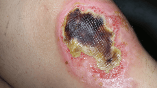
Burns: Clinic, diagnosis and treatment
Medical editor: Dr. Pierre Schneider, Dermatologist, Saint-Louis Hospital, France.
Related topics
Key messages:
- Burns are injuries caused by heat, cold, chemicals, electricity, radiation or mechanical energy.
- They are classified into several levels according to the depth of tissue damage. In the most severe cases, they may require hospitalization.
- Their classification depends on the extent of the lesions, their location and the type of lesions present.
- The first reflex in case of a superficial thermal burn is to leave the burned area under cold water for at least 5 minutes.
- Burns are tissue injuries caused by exposure to heat, cold, chemicals, electricity, radiation or mechanical energy.
- Burns can be classified into three categories based on their depth: first-degree burns, which affect only the epidermis; second-degree burns, which also affect the dermis; and third-degree burns, which affect the deep layers of the skin and subcutaneous tissue.
- Burns can cause severe pain, inflammation, loss of sensation, tissue necrosis and permanent damage.
- Treatment of burns depends on their severity and may include local care, surgical interventions, and rehabilitative therapies1,2.
The physiopathology of burns is complex and depends on the depth, extent and cause of the burn2,4:
First-degree burns
- Are burns that cause superficial damage to the epidermis, which is the top layer of the skin.
- The cells of the epidermis are damaged or destroyed, resulting in local inflammation and severe pain2.
Intermediate second-degree burns
- Cause deeper damage, reaching the dermis, the layer of skin beneath the epidermis. Blood vessels are damaged, causing plasma to leak and fluid to accumulate in the tissue.
- This can cause blistering, skin loss and increased pain.
- They can be separated into two subclasses2:
- The second superficial degree: it shows total involvement of the epidermis and the papillary dermis. The clinical sign is a blister, which then leaves a pink, oozing, painful erosion. Healing is spontaneous in less than 14 days, but residual dyschromia is possible.
- The second deep degree: it shows epidermal destruction with damage to the reticular dermis, but preservation of the appendages. The clinical sign is a blister with a reddish-brown background, leaving a whitish, atonic, hypoesthetic erosion with detachment of the cutaneous appendages. Healing is slow, taking 3 to 6 weeks, often at the cost of residual hypertrophic scars.
Distinguishing between a superficial and a deep second-degree burn can be difficult, especially since the burn may worsen over time2.
Third-degree burns
- Cause damage to the deep layers of the skin and subcutaneous tissue, resulting in loss of sensation and tissue necrosis.
- If smoke or fumes are inhaled, these burns can also cause damage to internal organs such as the lungs or the nervous system2.
Fourth degree burns or carbonization
- We speak of fourth degree burns or carbonization in case of fat or muscle damage.
- The clinical sign is the white or black aspect, somewhat cardboardy and anesthetized.
- No healing is possible except from the edges, which are often far apart.
- Surgical treatment is mandatory1,5.
The inflammatory response is a key element in the physiopathology of burns. Immune cells, such as neutrophils and macrophages, migrate to the burn area to remove dead cells and tissue debris. However, this excessive inflammatory response can cause damage to healthy tissue, resulting in a systemic response called systemic inflammatory response. Cytokines, signalling molecules involved in inflammation, are released from the damaged cells, and can cause disruptions in clotting, blood pressure, and organ function4.
In conclusion:
- Burn healing is a complex process that involves the regeneration of skin cells and the formation of collagen to replace damaged tissue.
- Second and third-degree burns can cause scarring and disturbances in skin pigmentation.
- Finally, functional and cosmetic sequelae may persist after burns heal4.
Acute Burn2
In the case of an acute burn, several factors must be determined:
Extent of injury
- Wallace's Rule of Nines is a tool used to assess the extent of body burns in adults. To use Wallace's Rule of Nines, the body is divided into nine equal sections and the percentage of burns in each section is calculated. Using this information, the physician can assess the severity of the burn and plan appropriate treatment.
- It is important to note that this rule is primarily used for adults and may not be accurate for children, obese or skinny people because their total body surface area may vary.
Depth of lesions
- Simple erythema: superficial first-degree burn.
- Erythema + blisters with exposed, red, moist but sensitive dermis: second-degree burn, superficial or intermediate.
- Insensitive dermis turned white or charred: third-degree burn.
Contributing factors
- Origin:
- Liquid.
- Solid.
- Gas.
- Electricity.
- Observations:
- Ensuring whether eyes were affected or not as it warrants an urgent consultation with an ophthalmologist.
- Burns present on sensitive areas (face, neck, genitals).
- Involvement of areas susceptible to functional sequelae (hands, folds, periorificial region).
- Circular burns on limbs.
- Terrain:
- Children - elderly patients – at-risk patients (heart failure, respiratory difficulties, renal function, alcoholism, diabetes).
- Septic conditions with major risks of superinfection.
- Commonly associated signs: signs of shock:
- Blood pressure.
- Temperature.
- Pulse.
- Other lesions (fractures).
Chronic Burn3
- Chronic burn, also known as erythema ab igne or hot water bottle rash, is a skin condition caused by prolonged exposure to heat.
- The symptoms typically include redness, itching, blistering and burning on areas of the skin exposed to heat.
- This condition is most common in people who work in hot environments, such as construction workers, furnace workers and kitchen workers. Excessive heat can cause blood vessels in the skin to dilate, resulting in redness and irritation3.
- Symptoms can be more severe if the skin is also exposed to irritants such as chemicals or abrasive substances.
- In some cases, hot water bottle rash can worsen and progress into a bacterial or fungal infection3.
- Preventative measures should be taken to avoid hot water bottle rash, such as wearing protective clothing to cover areas of the skin exposed to heat, using moisturizers to protect the skin, and taking regular breaks to cool down3.
Treatment of burns depends on the severity of the burn:
Mild burns
Can often be treated with home care such as placing a sterile dressing and continuing topical care to prevent infection and promote healing.
Moderate to severe burns
They usually require professional medical care2.
- First-degree (epidermal) burns are usually treated locally with the use of sterile dressings and analgesics to relieve pain.
- Second-degree (partially deep) burns usually require professional medical care and may require hospitalization for care such as surgical debridement of the burnt area, placement of special dressings to promote healing, and prescription of painkillers to relieve pain.
- Third-degree (deep) burns are the most severe and may require hospitalization, surgical procedures to remove burnt tissue, and skin grafts to promote healing2.
Good to know:
- It is important to closely monitor patients with severe burns to detect and treat potential complications such as renal failure, hypovolemia, infection, and smoke inhalation.
- This might require for patients to be treated in an intensive care setting1,5.
In case of acute burn
For superficial thermal burns
- First aid:
- The first step is to cool the burnt area under running cold water for at least 5 minutes (the cold temperature triggers a vasoconstriction that reduces the inflammation caused by the burn).
- Cleaning the area with water, soap or with an antiseptic alcohol-free solution.
- Never deroof a blister as its roof serves as a natural protection. If the blister is ruptured, disinfect and protect the area with a dressing appropriate to the size of the burn, possibly using a paraffin gauze dressing.
- Apply emollient such as tulle gras or specialist scarring products.
- Use topical antibiotics in case of superinfection (fusidic acid, mupirocin).
- Second step: application of compression bandages: siliconized patches that limit the risk of hypertrophic or keloid scars1,2.
For chemical burns
The acidic or basic solution should be neutralized by its antidote, or else by continuous irrigation for 10 min with tap water (except for burns inflicted by self-defence bombs)1,2.
Considerations
- Associated lesions (fractures).
- Patients representing a higher risk (heart failure, respiratory difficulties, renal function, alcoholism, diabetes).
- Elderly patients or children.
- Severe burns on hands.
- Burns of the respiratory or upper digestive tract (chemical fumes, gas or caustic liquid).
- Septic burns.
- Widespread and deep burns.
- Burnt surface area over 10 to 15 % on an adult, 5 to 10 % on a child.
Then hospitalization in a specialist Burns Service center. The main principles of care are1,5:
- Excision of necrotic tissues in one or more steps.
- Compensations of caloric and thermal losses.
- Screening and treatment of superinfections.
- Autologous thin skin grafts from intact areas or obtained by epidermal culture from a small fragment of the patient's healthy skin taken upon admission.
- Extended physiotherapy.
- Compression garments for hypertrophic scars.
- Postural splints.
- Thermal cures with filiform douches, etc.
In case of chronic burn
No treatment needed: ending contact allows a progressive regression of the symptoms3.
How are burns on a pregnant woman treated?
- Treatment must be adapted to the severity of the burns.
For a pregnant woman: are recent scars in the epidural injection area a problem?
- Not if the wound is closed; epidural injection is not recommended in the presence of open wounds.
Are there any treatments able to help the recovery of sensitivity following a burn?
- In some cases, physiotherapy and massages can help recover sensitivity.
What does the presence of telangiectasias mean?
- Telangiectasias can be associated with a specific type of burns such as radiation burns.
- Ayar et Benyamina, Prise en charge du patient brulé en préhospitalier. Première partie : cas général et inhalation de fumées, Annals of burns and fire disasters, volXXXII – n.1 – Mar 2019
- Ingen Housz Oro et al, Brûlures superficielles : physiopathologie,clinique, traitement. Doi : 10.1016/S1634-6939(10)55123-8
- Miller et al, Erythema ab igne, Dermatol Online J. 2011 Oct 15;17(10):28.
- Roshangar et al, Skin Burns: Review of Molecular Mechanisms and Therapeutic Approaches. Wounds. 2019 Dec;31(12):308-315.
- Brûlures de la peau. Ameli. https://www.ameli.fr/assure/sante/urgence/accidents-domestiques/brulures-peau, website consulted on 30/01/2023
Create easily your professional account
I create my account-
Access exclusive business services unlimited
-
Access valuable features : audio listening & tools sharing with your patients
-
Access more than 150 product sheets, dedicated to professionals


